Week 4!
Thought I would lead with a picture of what I’ve been doing lately:
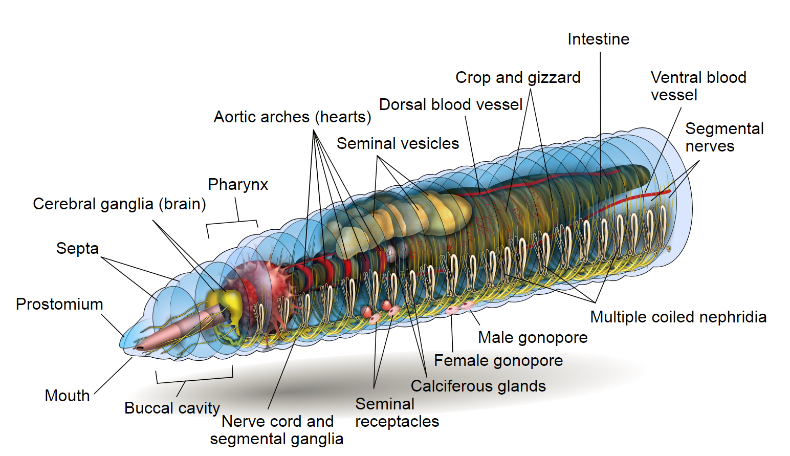
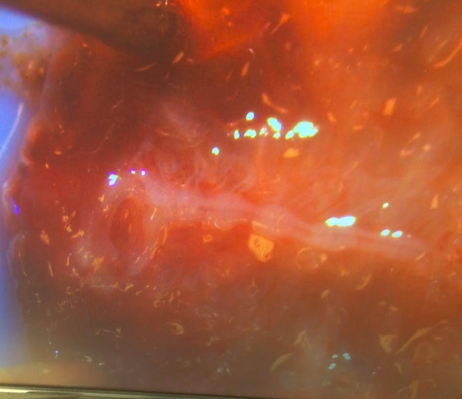
The horseshoe attached to the white line is the brain (two lobes) and the nerve cord of one of the earthworms. We’ve been trying to develop an efficient method for exposing the brain so that when we fix the samples and introduce neuropeptide antibodies onto them, binding can occur. If you look at the diagram, the brain and mouth actually flip dorsal and ventral sides so that the gut lies above the nerve cord, which makes it tricky to get to the brain.
I’ve also been practicing the other set of dissections for DASPEI staining but those weren’t with a microscope hooked up to a monitor so the pictures aren’t large enough to show anything noticeable.
Working in a Biology lab has brought to mind the phrase, “Hurry up and wait”, as there is often down time as you wait for the next steps in a procedure to finish. Practicing dissections under a microscope has also taught me the difference between practicing until you can get it right and practicing until you can’t get it wrong. This has been a valuable difference to keep in mind so far and I’m sure it will continue to apply in the future.
ohari15 • June 13, 2018